

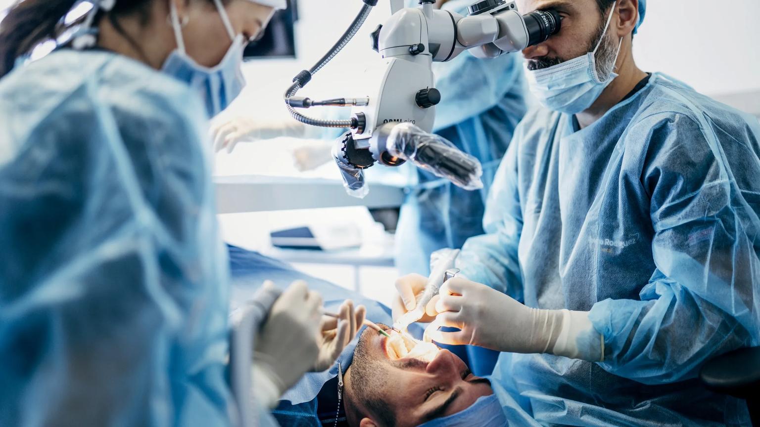
What are the ten regions of full-face and neck toxin treatment?
In the masterclass's framework the ten regions are the glabellar complex, the frontalis, the periorbital area, bunny lines, the gummy smile, perioral lines, marionette lines, the mentalis, the masseter and the platysmal bands. Each is defined by the muscle it targets rather than by the line it treats, which is why the anatomy, not the indication, sets the depth.
The table groups the ten into six teaching blocks, in the order the hands-on block works down the face on the first afternoon, and lists for each the target muscle, the depth that matters, the neighbour behind the recognised complication and what the dissection shows.
| Block | Target muscle | Depth that matters | Neighbour to respect | What dissection shows |
|---|---|---|---|---|
| Glabellar complex | Procerus and corrugator supercilii | Corrugator deep at its bony origin, superficial towards its insertion | Levator palpebrae superioris (eyelid ptosis) | The corrugator's course from deep origin to superficial insertion |
| Frontalis and periorbital area | Frontalis; orbicularis oculi | Frontalis a thin sheet under the skin; orbicularis superficial over the orbital rim | Lower frontalis (brow ptosis); zygomaticus major (smile asymmetry) | How thin the frontalis is and where the orbicularis meets the cheek muscles |
| Bunny lines and gummy smile | Nasalis; levator labii superioris alaeque nasi | Nasalis superficial over the nasal bones; the levator within the muscle beside the ala | Levator labii superioris (upper-lip ptosis) | The levator's course from the medial orbit along the nose to the ala and lip |
| Perioral and marionette lines | Orbicularis oris; depressor anguli oris | Superficial, small volumes | Depressor labii inferioris (lip ptosis); the modiolus | The modiolus as the muscular crossroads of the mouth |
| Mentalis | Mentalis | Deep, at the chin | Depressor labii inferioris | The mentalis origin on bone and its upward fibres |
| Masseter and platysmal bands | Masseter; platysma | Masseter deep and thick; platysma thin and superficial | Risorius and zygomaticus (smile); the structures beneath the platysma | Masseter thickness; the platysma as a sheet over the neck |
Why is depth the variable that matters most for toxin?
Because the muscles differ in depth and thickness far more than diagrams suggest: the frontalis is a thin sheet, the corrugator runs from deep origin to superficial insertion, the mentalis sits on bone, the masseter is thick and deep. The Global Aesthetics Consensus Group's recommendations on botulinum toxin type A (2016) base injection dosage and placement on analysis of the target muscle in the context of adjacent muscles and the associated soft and hard tissues, and note intracutaneous injection where indicated to limit the depth and degree of action.
Toxin diffuses from the point of injection, so a dose placed one layer too superficial or too deep reaches a neighbouring muscle rather than the target. That is the mechanism behind the predictable complications: brow ptosis when the frontalis is treated too low, lip ptosis when the depressor labii inferioris is caught instead of the depressor anguli oris, an asymmetric smile when the zygomaticus or risorius is reached from a periorbital or masseter point.
Which neighbours cause the complications injectors fear?
The levator palpebrae superioris behind the glabellar complex, the lower frontalis above the brow, the depressor labii inferioris beside the depressor anguli oris, the zygomaticus and risorius near the periorbital and masseter points, and the structures beneath the platysma. Every one of them is a structure an injector cannot see from the surface and that a dissection exposes in minutes.
The lower face and neck carry the least familiar neighbours. A cadaveric study of the cervical nerve for face and neck lifting, co-authored by the course director and published in Aesthetic Surgery Journal (2024), is an example of how much runs under the platysma that a platysmal-band injection has to respect.
What does a cadaver dissection add for toxin, compared with filler?
For filler, dissection verifies the plane a dyed product reached; for toxin, dissection shows the muscle itself: its thickness, its borders, its depth under the skin and its relationship to the neighbour behind the complication. In the masterclass the toxin block is hands-on on the fresh-frozen specimen after the day's two dissection sessions and the instructor's demonstration of muscle depths and injection points on the cadaver; dye verification is the day-two method for filler and biostimulator placement.
Estai and Bunt's review of anatomy teaching practice (2016) keeps cadaveric dissection as the reference method because it couples the three-dimensional specimen with immediate correction; for toxin, the spatial relationship between the target and its neighbour is the whole skill.
How does the masterclass sequence the ten regions?
On the first afternoon, after the theory session and the two instructor-led dissection sessions: a demonstration, then hands-on practice by every participant on all ten regions. The demonstration covers pharmacology, reconstitution and dosing strategy, with muscle depths, injection techniques and injection points shown on the cadaver by the instructor physician; the hands-on block then runs from the glabellar complex to the platysmal bands. Between three and six participants share one fresh-frozen cadaver head. The programme facts for the ATDERA edition of March 2027 are tabulated below. ATDERA is the organiser and coordinator of the masterclass; the course director and faculty deliver the teaching in an anatomy laboratory in Ankara; where a credential is involved, the accrediting body is a separate third party — and for this masterclass no accreditation is claimed.
| Field | Detail | Where to verify |
|---|---|---|
| Dates and venue | Saturday 27 and Sunday 28 March 2027; TORLAK & TunaliS Anatomy Institute Joint Lab, Ankara | Programme page: hero and laboratory section |
| Language and group | English; up to six participants; between three and six participants share one fresh-frozen cadaver head | Programme page: at a glance |
| Eligibility | Licensed physicians; foundation-level injectables experience recommended; international registrations welcome; every application reviewed academically | Programme page: who should apply |
| Fee and what it covers | £2,100 per participant, collected in GBP after written acceptance; both laboratory days, dyed training products and consumables, personal protective equipment, lunch and coffee breaks; accommodation and transfers available separately on request | Programme page: fee section |
| Certificate and CPD | ATDERA certificate of attendance on attendance across both days and participation in every hands-on block; no CPD or CME credit is claimed | Programme page: certificate line and FAQ |
| Organiser and verification | ATDERA Global Limited, registered in England and Wales, Companies House number 17173428; ATDERA organises and coordinates, the course director and faculty deliver the teaching | Companies House register; programme page footer |
What should an injector check before treating a new region?
Five things, in the order the anatomy imposes them.
- Which muscle is the target, and is it a sheet, a band or a thick mass?
- At what depth does the target sit under the skin at the chosen point, and does that change along its length?
- Which neighbour lies within the diffusion radius, and what does it do if it is reached?
- What does the consensus guidance say about points and depth for this region?
- Have you seen the muscle exposed on tissue, or only drawn?