Why bone grafting exists in implant dentistry
Dental implants depend on a critical input: enough living bone at the planned implant site to support primary stability of the implant at placement and to support direct bone-to-implant integration in the months that follow. Where that bone volume is present, the implant is placed directly. Where it is reduced — by long-term tooth loss, periodontal disease, traumatic loss, or pneumatisation of the maxillary sinus into the upper jaw — the planned implant either cannot be placed at all or cannot be placed at the dimensions required for long-term stability. Bone grafting exists to bridge that gap.
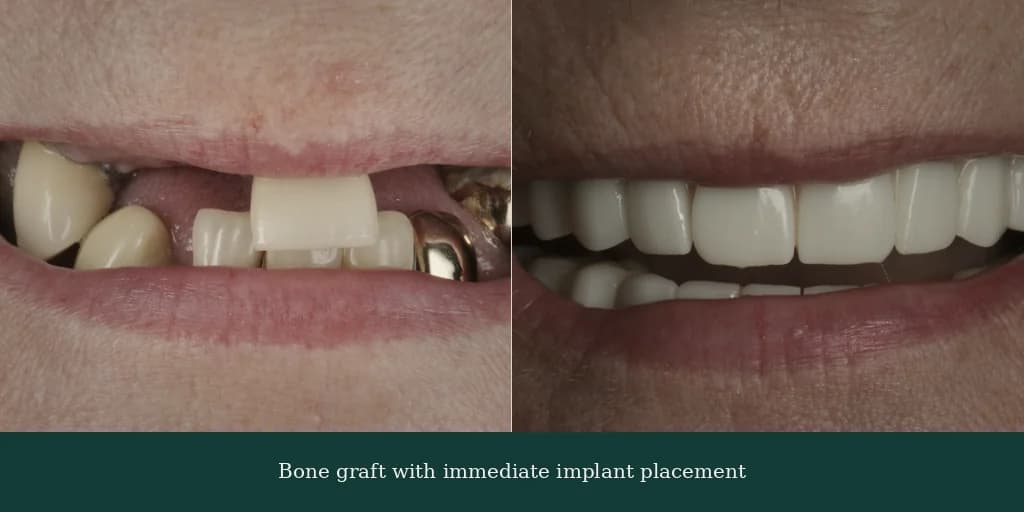
Grafting is not a separate clinical aim from implant treatment. It is an adjunctive procedure that creates the structural conditions for a successful implant placement. Where grafting is required, it is planned alongside the implant pathway from the outset rather than as a surprise step encountered after the patient has committed to treatment. The CBCT scan is the diagnostic that decides whether grafting is needed, what type, where, and in what volume; the verified partner implantologist's reading of the scan produces a written augmentation plan before any travel plan is made.
A patient who has been told elsewhere that they are not a candidate for implants because of insufficient bone is in many cases a candidate after a structured bone augmentation plan is in place. The ATDERA pathway exists in part because bone augmentation is a routine pre-implant procedure with well-established clinical outcomes when performed by a trained implantologist or maxillofacial surgeon, and a second-opinion remote case review of the CBCT scan is the appropriate first step where the original assessment ruled the patient out without explicit reference to the augmentation options below.
How bone is lost — the biology behind alveolar resorption after tooth extraction
Alveolar bone is the segment of jawbone that surrounds and supports the roots of the natural teeth. Its presence and structure depend on the functional load that the teeth transmit through the periodontal ligament — the bone is maintained by the mechanical signal of chewing. When a tooth is lost, that mechanical signal is removed, and the alveolar bone begins to resorb. The resorption is most rapid in the first six to twelve months after extraction and then continues at a slower rate over the years and decades that follow.
The pattern of resorption differs between the upper and lower jaws and between anterior and posterior regions. In the upper jaw, posterior resorption combines vertical bone loss in the alveolar ridge with downward expansion of the maxillary sinus into the space the bone previously occupied — a process termed sinus pneumatisation. In the lower jaw, resorption is more uniform but tends to reduce ridge width before ridge height, which can compromise implant placement even where the radiographic ridge appears to retain reasonable height. Both patterns are surfaced by CBCT imaging and inform the augmentation plan.
Long-term denture wearers and patients with longstanding partial edentulism are the two clinical groups in whom alveolar resorption is most extensive. Periodontal bone loss adds a second mechanism — bacterial inflammation breaks down the bony support around remaining teeth and leaves residual ridge defects after extraction. Traumatic tooth loss, particularly with associated alveolar fracture, can produce localised defects that are not predicted by the timeline of tooth loss alone. Each of these mechanisms is read on the CBCT scan, and the augmentation plan responds to the specific defect pattern rather than to a generic time-since-extraction estimate.
The four graft material categories — autograft, allograft, xenograft, and alloplast
Bone graft materials are classified into four clinical categories, each with a different biological profile and a different set of indications. Autograft is the patient's own bone, harvested from a secondary site — most commonly the chin, the mandibular ramus, or, for larger volumes, the iliac crest or tibia. Autograft contains the patient's own osteogenic cells and growth factors and is the most biologically active option, but it requires a second surgical field and donor-site morbidity, which limits routine use to cases where biological activity is the dominant consideration.
Allograft is processed donor bone from a regulated tissue bank — human cadaveric bone that has been screened, decellularised, and sterilised to remove infectious risk while retaining the bone's mineral structure and, in some preparations, demineralised bone matrix that supports new bone formation. Allograft is widely used in routine grafting because it provides reliable structural performance without a secondary surgical field for the patient, and it is supplied to the surgical site through internationally regulated tissue bank channels.
Xenograft is bovine-derived deproteinised bone mineral — most commonly Bio-Oss (Geistlich) and equivalent products from other manufacturers — in which the organic component of bovine bone has been removed and the remaining hydroxyapatite mineral matrix acts as a scaffold for new bone formation by the patient's own cells. Xenograft is the most commonly used graft material in routine maxillary sinus lift surgery and ridge augmentation because of its predictable resorption and remodelling profile, and because it is widely supported by long-term clinical literature.
Alloplast is a synthetic biocompatible substitute — typically a calcium phosphate or beta-tricalcium phosphate ceramic, a bioactive glass, or a synthetic hydroxyapatite. Alloplast carries no biological transmission risk, is structurally consistent batch to batch, and is increasingly used as a primary or supplementary graft material in routine cases. The choice between the four categories is made by the verified partner implantologist or maxillofacial surgeon based on the volume required, the surgical site, the planned implant timeline, and the patient's clinical and personal preferences. The selection is recorded in the case-specific written estimate before surgery.
Sinus lift surgery for posterior maxillary implants
The maxillary sinus is the air-filled cavity in the upper jaw, located above the roots of the posterior maxillary teeth. After loss of upper-jaw posterior teeth, the maxillary sinus tends to expand downward into the space the alveolar bone previously occupied, reducing the residual sub-antral bone height available for implant placement. Where that residual height is below the threshold for direct implant placement — typically below five millimetres for a standard-length implant — a maxillary sinus lift is the appropriate adjunctive procedure.
Two surgical techniques are used. The lateral window approach (also termed the Caldwell-Luc approach) creates an access window in the lateral wall of the maxillary sinus, elevates the sinus membrane carefully off the bony floor, and packs graft material below the elevated membrane to create new sub-antral bone height. The crestal approach (also termed the osteotome or Summers technique) elevates the sinus membrane from below through the planned implant osteotomy, packs a smaller volume of graft material, and is appropriate where the residual bone height is in the moderate range and the volume of augmentation needed is modest. The verified partner clinician selects the appropriate technique on the basis of the CBCT scan and documents the rationale in the written augmentation plan.
Long-term clinical literature reports favourable outcomes for maxillary sinus floor elevation. Published systematic reviews report implant survival above 90% at five years for implants placed into sinus-lift-augmented sites, and the procedure is supported by a substantial body of peer-reviewed clinical research dating back to the 1980s. The most commonly reported intra-operative finding is small membrane perforation, which is managed at the time with a barrier membrane and rarely affects the final outcome. Post-operative sinus inflammation or, less commonly, sinus infection is monitored through the structured remote review schedule the partner clinician puts in place after surgery.
Ridge augmentation for narrow ridges
Ridge augmentation is the second of the two principal pre-implant grafting procedures. It addresses width or height loss at the alveolar ridge — most commonly in patients with longstanding tooth loss in the lower jaw or in the anterior maxilla, where vertical resorption and lateral resorption produce a ridge that is too narrow to support a standard-diameter implant or too short to support an implant of adequate length. The augmentation places graft material against the residual ridge and contains it under a barrier membrane while new bone forms around and through it.
Two protocols are commonly used. Guided bone regeneration (GBR) uses a particulate graft (xenograft, allograft, or alloplast) under a resorbable or non-resorbable barrier membrane to direct new bone formation into the augmented site while excluding soft tissue from invading the graft volume. Block grafting uses a structurally intact piece of bone — most commonly autograft from a donor site, sometimes processed allograft block — fixed to the residual ridge with internal screws and covered with a particulate graft and barrier membrane to support remodelling. The choice between GBR and block grafting depends on the volume of augmentation required, the surgical site, and the verified partner clinician's reading of the CBCT scan.
Recovery from ridge augmentation follows the broader bone-graft pattern: soft-tissue healing in one to two weeks, bone consolidation over three to six months for staged protocols, and a CBCT review at the end of consolidation to confirm bone height and density before staged implant placement. Patients with larger augmentation volumes or with augmentation in challenging surgical sites (for example, a severely resorbed anterior maxilla) may require a longer consolidation period, sometimes up to nine months, before the implant placement visit. The timeline is documented in writing in the case-specific written estimate before surgery proceeds.
Staged vs simultaneous protocol — when graft and implant are placed together vs separately
A central planning decision in any bone-grafting case is whether the implant is placed at the same surgical visit as the graft, or whether the graft consolidates first and the implant is placed at a second surgical visit three to six months later. The decision depends on whether the residual bone volume is sufficient to support primary implant stability at the time of grafting. Where it is — typically a sinus lift with adequate pre-existing sub-antral bone, or a localised ridge graft of modest volume — the simultaneous protocol is feasible and shortens the overall pathway. Where the residual bone volume is more limited, the staged protocol is the more predictable plan.
The staged protocol separates the bone augmentation and the implant placement into two distinct surgical events. The first visit covers the graft, with three to six months of consolidation following. A CBCT review at the end of the consolidation period confirms bone height and density, and the implant is then placed at the second surgical visit. The two-stage timeline adds three to six months to the total pathway but produces more predictable primary stability of the implant in cases where the augmentation volume is substantial. The full pathway from initial graft to final prosthesis can extend to nine to twelve months in staged cases.
The simultaneous protocol places the graft and the implant in the same surgical session. The graft material is positioned around or alongside the implant at placement, often under a barrier membrane, and the implant integrates into the consolidating graft volume during the standard three-to-six-month osseointegration window. The simultaneous protocol shortens the overall pathway and avoids a second surgical visit, but it requires sufficient native bone for the implant to achieve primary stability at the time of placement. Where primary stability is borderline, the verified partner clinician converts to the staged protocol intra-operatively rather than placing the implant under suboptimal conditions.
The decision between staged and simultaneous is made on the CBCT scan and the verified partner clinician's reading of the residual bone volume. The rationale is documented in writing in the case-specific written estimate before any travel plan is made, so the patient and the patient's UK or home-country dentist can read why one protocol is recommended rather than the other. Patients sometimes ask whether the simultaneous protocol can be requested to shorten the pathway; where the clinical situation supports it the protocol is already the recommendation, and where it does not the clinical risk of compromised primary stability is documented in the written summary rather than reframed as a patient preference.
Recovery timeline and the wait before implant placement (3 to 6 months staged)
Recovery from a bone graft or sinus lift follows a documented pattern. Soft-tissue healing typically takes one to two weeks, during which mild swelling around the surgical site is managed with prescribed analgesics, a chlorhexidine rinse protocol, and a soft diet. For sinus lift cases, additional precautions apply: patients avoid blowing their nose forcefully, avoid air travel for the first two weeks where possible, and avoid activities that increase pressure in the sinus cavity (deep diving, vigorous bracing, instruments that apply intra-oral pressure). The verified partner clinician confirms the appropriate restriction window at the in-country review.
Bone consolidation — the biological remodelling of the graft into the patient's native bone structure — takes three to six months in most cases. During this period, the graft material is gradually resorbed and replaced by newly formed bone in continuity with the surrounding native bone. The process is not visible to the patient and proceeds in the absence of observable change at the surgical site, which mirrors the quiet phase of osseointegration in implant treatment more broadly. Patients sometimes find this period harder to live through psychologically than the immediate post-operative window, which is why the structured remote review schedule (week 1, week 6, month 3) provides documented confirmation that consolidation is proceeding as expected.
At the end of the consolidation period, a CBCT review confirms bone height and density at the augmented site. Where the augmentation has consolidated to the planned volume, the implant placement visit is scheduled. Where consolidation is partial or delayed — sometimes encountered in larger augmentation volumes or in patients with slower bone-healing biology — the wait is extended by two to three months and a follow-up CBCT confirms readiness before the implant placement visit. The decision to extend the wait is a clinical decision rather than a scheduling preference, and it is documented in writing.
When the implant is placed at the augmented site — whether at the staged second visit or simultaneously with the graft — the placement is recorded on the manufacturer's implant passport. Straumann's Patient Pass, Nobel Biocare's Implant Passport, or the equivalent documentation for the implant system used captures the implant brand, the abutment configuration, the surgical date, the treating clinician, and the implant serial numbers. The patient travels home with the passport as the technical record of the augmented-site implant, and ATDERA retains a duplicate in the pathway file.
The alternatives — when bone grafting can be avoided
Bone grafting is not the only response to insufficient residual bone. Three alternative techniques are routinely considered, and the verified partner clinician on the ATDERA pathway documents the rationale for grafting versus an alternative in the written case plan. The first alternative is the angled posterior implant — most often used in full-arch All-on-4 protocols, where the two posterior implants are angled by up to 45 degrees to maximise contact with available bone, often allowing treatment to proceed without a separate sinus lift or extensive grafting procedure.
The second alternative is the short implant — a standard-design implant of reduced length (typically six to eight millimetres rather than ten to thirteen millimetres) placed where the residual bone height is reduced but the bone density is sufficient to support primary stability of the shorter implant. Short implants are supported by long-term clinical literature in selected indications and are most commonly considered in the posterior mandible where vertical bone height is reduced by proximity to the inferior alveolar nerve canal. The verified partner clinician confirms whether a short implant is the appropriate response to the specific bone profile, and the choice is documented in writing.
The third alternative is the zygomatic implant — extra-long titanium implants anchored not in the alveolar bone of the maxilla but in the zygomatic (cheek) bone above and lateral to the upper jaw. Zygomatic implants are used in cases of severely resorbed maxilla where bone grafting is contraindicated, has previously failed, or has been declined by the patient. The technique was established by the Brånemark zygoma protocol and is now supported by long-term clinical evidence, and a separate ATDERA-coordinated pathway exists specifically for patients with severely resorbed maxilla who have been told elsewhere that they have no fixed-prosthesis option without grafting.
The clinical decision between bone grafting and one of the three alternatives is not a tier selection or a preference question. It is a structural decision driven by the residual bone volume on the CBCT scan, the patient's medical history, and the prosthetic plan. The verified partner clinician on the ATDERA pathway maps the case onto the appropriate option at the remote case review, and the rationale is recorded in the case-specific written estimate so the patient and the patient's UK or home-country dentist can read why one path is recommended rather than another.